background

Retina Imaging

Retina Imaging

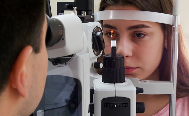
Retina imaging is a branch of medical imaging focused on capturing detailed images of the retina, the light-sensitive tissue lining the back of the eye. The retina plays a crucial role in vision by converting light into neural signals that the brain interprets as images. Imaging techniques help in diagnosing and monitoring various eye conditions, including diabetic retinopathy, age-related macular degeneration, glaucoma, and retinal detachment. Best Retina Specialist in Ghatkopar

These imaging modalities play a crucial role in the diagnosis, management, and monitoring of retinal diseases and conditions. They allow eye care professionals to visualize changes in the retina over time, track disease progression, and make informed decisions about treatment options.

Common techniques used in retina imaging include:

  • Fundus Photography : This involves capturing high-resolution images of the retina using a specialized camera called a fundus camera. It helps in documenting the appearance of the retina, optic nerve, and blood vessels.
  • Optical Coherence Tomography (OCT) : OCT is a non-invasive imaging technique that uses light waves to produce high-resolution cross-sectional images of the Best Retina Specialist in Ghatkopar. It provides detailed information about the layers of the retina, allowing for the detection of abnormalities such as swelling, fluid accumulation, or thinning.
  • Fluorescein Angiography (FA) : FA involves injecting a fluorescent dye into the bloodstream, which highlights the blood vessels in the retina. Images are then captured as the dye circulates through the blood vessels, helping in the diagnosis of conditions such as macular degeneration, diabetic retinopathy, and retinal vascular occlusions.
  • Indocyanine Green Angiography (ICG) : Similar to FA, ICG uses a dye to visualize blood vessels, but it provides deeper penetration and is particularly useful for imaging the choroid, the vascular layer beneath the retina.
  • Scanning Laser Ophthalmoscopy (SLO) : SLO produces high-resolution images of the retina using a scanning laser beam. It enables the visualization of individual retinal cells and is often used in research and advanced diagnostic procedures.
  • Multifocal Electroretinography (mfERG) : mfERG measures the electrical responses of different areas of the retina to light stimulation. It helps in evaluating the function of specific retinal areas and is particularly useful in diagnosing conditions affecting the central vision.
  • Important Link